What Are the Many Ways to Remove Earwax

What Are the Many Ways to Remove Earwax
There are four popular methods for removing earwax. These are earwax drops, ear syringes or irrigation, micro-suction earwax removal, and endoscopic earwax removal.
Drops of Earwax
Any pharmacy will have a variety of over-the-counter medicines that will loosen, soften, or dissolve earwax. These might be oil-based (olive oil-based) or water-based (sodium bicarbonate or saline). Water-based drops are more effective than oil-based solutions for dissolving earwax. If perforation has previously been diagnosed, the drops should be avoided because they can cause difficulties with middle ear infections. The drops are administered at home and must be kept at room temperature to avoid the “caloric effect,” which occurs when the ear balance organ is out of rhythm. This is due to cooler air or water inhibiting its function.
Applying earwax drops can be both painful and time-consuming. When applying the drops, the affected ear must be turned up. After application, you must remain in place for 5 to 10 minutes before sitting up and clearing away any excess ear wax. This method must be repeated 2/3 of the time per day for 10 to 14 days. Even after this surgery, using drops alone may not be effective if the earwax is impacted.
Irrigating the Ears
A medical assistant, a community nurse, and some audiologists typically do ear irrigation. It is usually provided free of charge by the NHS. A metal ear syringe was traditionally filled with warm water, and the metal tip was placed into the ear canal. The water was then injected into the ear canal, and a kidney dish was placed behind the ear to collect the water and earwax that was washed out. The syringe must be lubricated regularly to apply consistent pressure. Previously, the nurse would use their judgement to choose how hard to spray the water. An ear irrigation pump has replaced the metal ear syringe with a nozzle tip for safety reasons. Although the pump has a changeable, regulated pressure, the procedure remains fundamentally the same. Many people have had the ear syringe or irrigation administered multiple times without incident.

Side Effects, Hazards, and Disadvantages
When it works, it works well. However, there are several risks and downsides of ear syringing:
Before injecting, hard earwax must be softened for up to two weeks.
If the angle of the jet is not calibrated correctly, injecting can push wax deeper into the ear.
Tinnitus may result.
The eardrum may perforate.
Due to the abundance of earwax, an undiagnosed perforated eardrum may not be visible. Water, germs, earwax, and dead skin cells are flushed into the middle ear. This has the potential to result in a painful infection.
It is not advised to use after ear surgery.
Because of the risk of re-perforation, this procedure should not be undertaken if the eardrum has previously been perforated.
Under no circumstances should ear syringes be used on anyone with a known perforation, cleft palate, foreign body in the ear canal, or mastoid cavity following a mastoidectomy.
Many physician offices are discontinuing the use of ear syringes and directing all patients to the NHS ENT department. The internet site
There could be a considerable wait at the NHS ENT clinic.
Micro Suction Is Used to Remove Earwax
ENT doctors and nurses frequently do micro suction in hospital outpatient clinics when other procedures have failed. The main benefit of micro-suction is that earwax is always safely removed under direct view. The treatment is carried out in a hospital setting using a binocular ENT microscope to visualise the wax. After that, a sterile suction probe coupled with gentle suction equipment is employed. Other ENT instruments may be utilised as well. Microaspiration, unlike rinsing or spraying, is a “dry” process that does not require water. This dramatically minimises the chance of eardrum injury and infection.
What Is the Procedure’s Procedure?
Microaspiration is faster and safer, and it eliminates the need for weeks of ear drops. Microaspiration is done with giant, expensive binocular ENT surgical microscopes in ENT departments. Due to waiting lists, many people must wait weeks or months for an appointment, due to waiting lists. Some hearing care practitioners are now trained to perform a microaspiration, but they do so using microscope “loupes.” These resemble spectacles with headlights and are widely available and reasonably priced. They are not, however, as effective or as safe to use as ENT microscopes.
Risks and Side Effects
Noise may occasionally occur during this process due to the suction equipment. Dry skin flaps can vibrate, creating “carpeting.” Like ear syringing, some persons report tinnitus following microaspiration or an increase in tinnitus if they already had it before. Abrasions or scrapes in the ear canal caused by the suction probe pose a negligible risk of bleeding. Secondary eardrum injury from the suction probe and other ENT equipment is also conceivable but extremely rare. A caloric effect may arise during microaspiration due to the cooling effect of the suction probe. This may cause the patient to feel dizzy for a few minutes before the temperature returns to normal.
Earwax Removal through Endoscopy
Earwax removal
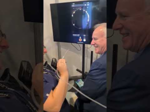
Endoscopic earwax removal, which is faster and easier to conduct than microaspiration, is an alternative. Endoscopic earwax removal is performed with an oto-endoscope. This is a more modern medical ear lamp than the typical otoscope used to check the ear by audiologists and physicians. It’s a small-diameter, rigid fibre-optic telescope that gives you an unrivalled “wide” and high-resolution view into your ear canal. It is both comfortable and safe because it can be retained in the ear canal and provides a view of the whole ear canal and eardrum with no manipulation of the patient’s head. On the other hand, the suction probe is used to remove earwax or implant additional ENT tools gently.
Endoscopic cerumen removal is preferable to microscopic cerumen removal, according to AJ Clegg et al. 2010 .’s Health Technology Assessment. Microscopic cerumen removal requires using a surgical microscope or surgical loupe to obtain a magnified view of the ear canal and eardrum – a speculum (the small funnel) must be inserted into the ear canal to provide a view. The speculum occupies some of the space in the ear canal, limiting the physician’s view even more. Furthermore, if the ear canal is particularly narrow, the speculum can occasionally cause minor damage to the skin in the ear canal.
What Is the Procedure’s Procedure?
On the other hand, endoscopic earwax removal entails introducing an exemplary 2.8-mm endoscope partially into the ear canal. The endoscope is a rod with fibre optics that transmit light from a light source to illuminate the ear canal and a fixed lens (or set of lenses) that conveys the vision from within to outside the ear canal. The endoscope can be examined directly or with an imaging device connected for better magnification.
Risks and Side Effects
Endoscopic earwax removal, like microaspiration, can be noisy. As with all previous earwax removal procedures, there is a minimal chance that some patients will experience tinnitus or an increase in tinnitus if they already had it before earwax removal. There is a slight chance of mild bleeding due to the suction probe scraping and scratching the ear canal. However, this risk is minimised compared to micro-suction because more working space is accessible. Secondary tympanic membrane injuries from oto-endoscopes, suction probes, and other ENT equipment are also uncommon. The “caloric effect,” as with micro-suction, is possible due to the cooling air currents generated by the suction machine, which might provide a brief feeling of dizziness that subsides after a few minutes.


